Being a lecturer in medical and forensic art has meant I’ve been involved in some fascinating design projects over the last 10 years.
My focus is on the future of medical art and artists, particularly in relation to new and developing technologies, and I’m also involved in projects and collaborations with many other members of staff and students. I have been involved in recreating faces of people who lived long ago – one of the most recent being Thankerton man, a copper age individual, for the Biggar & Upper Clydesdale Museum. This project involved creating a surface scan of the skull before undertaking a digital reconstruction process. The end result was 3D print of the reconstruction complete with glass eyes and a wig.
During 2016 I was lucky enough to lead on an exciting project involving the digitisation of specimens from the University of Dundee’s Zoology Museum. The Zoology Museum houses many fascinating specimens from around the world including the sectioned skull a Thylacine, otherwise known as the Tasmanian Wolf, an extinct species of carnivorous marsupial.
Most of the exhibits were collected by the celebrated Sir D’Arcy Wentworth Thompson, the first Professor of Biology at Dundee. 2017 is the centenary of D’Arcy Thompson’s influential book On Growth and Form and a number of events will be taking place around the country to celebrate.
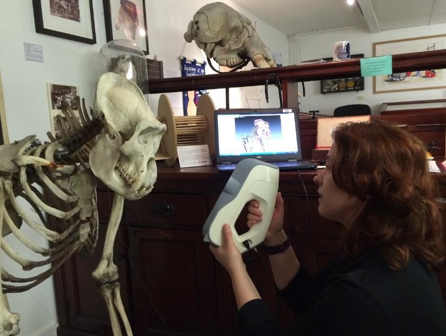
I became involved in the project after visiting the museum with the MSc Medical Art students. The Centre for Anatomy and Human Identification (CAHID) where I work had recently purchased two new structured light scanners (Artec Eva and Artec Space Spider) and I was keen to put them to the test. I had also recently been trained to use the University’s new micro CT scanner. The project seemed the perfect opportunity to hone my 3D scanning and modelling design skills.
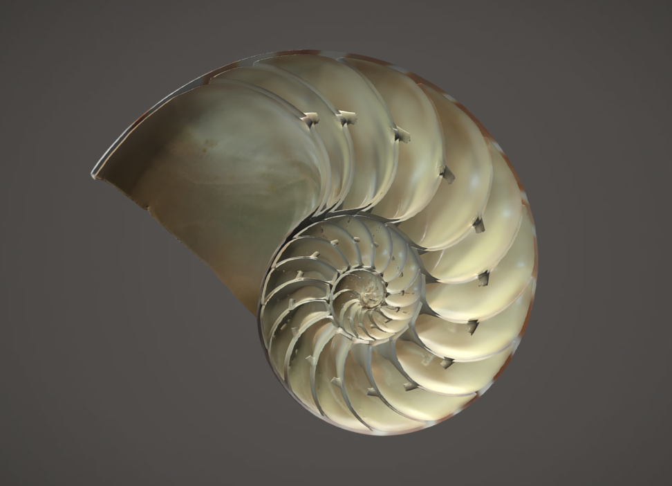
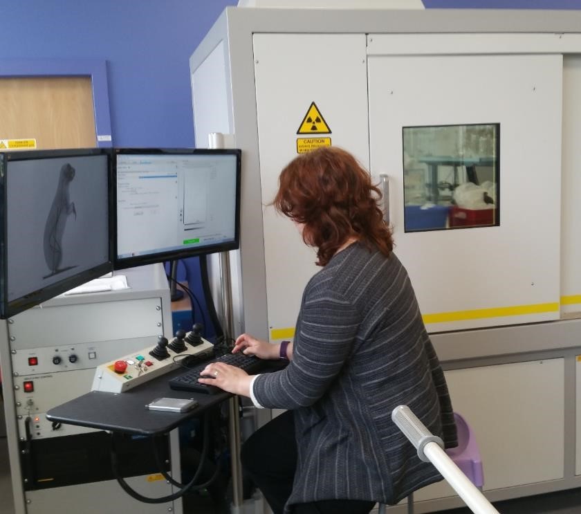
I worked on the project throughout 2016 whenever the opportunity arose between teaching and completing my PhD. Smaller specimens were scanned with the micro CT scanner, while larger specimens (from around 20cm) were captured using hand-held structured light scanners. An advantage of the micro CT scans was that they captured both internal as well as external surfaces, enabling them to be segmented, as in the example of the nautilus shell. The disadvantage however is that no colour information is captured. So all the colour had to be added later in the process.
The resulting 3D models are hosted online via Sketchfab and are available for viewing and downloading worldwide under a creative commons licence. Sketchfab is a website used to display and share 3D models online. They support cultural heritage institutions by providing free accounts and over 500 cultural institutions have joined Sketchfab so far. Once hosted on Sketchfab models can easily be embedded on websites and social media and even downloaded for 3D printing.
Finally, a webpage was created to showcase the models. The landing page features thumbnails of all available scans. When these are clicked on the user is taken to a page containing the 3D model and information about the specimen. The site can be viewed at: https://www.dundee.ac.uk/museum/collections/zoology/zoology3d/
The digital collection is being used in a variety of learning and teaching initiatives including by the University of Queensland who are incorporating the models into their level three vertebrate anatomy and evolution module, and the organisation ‘Museum in a Box’ who will be including one of the skulls in their school outreach program, where museum specimens are 3D printed and taken in to the classroom (http://museuminabox.org/).
The internet has been likened to the printing press of our time – it has disrupted the way the world creates, shares and consumes information. So far the models have been viewed over 8,000 times in more than 25 countries with over 1,500 downloads having taken place. This project aims to raise awareness of the collection, both locally and internationally, as well as provide access to those who may not otherwise be able to view it.
Dr Caroline Erolin is course co-ordinator for MSc Medical Art at the Centre for Anatomy and Human Identification.
