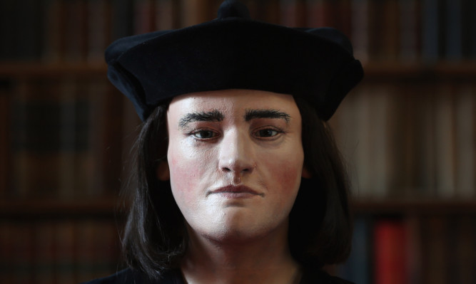
Dundee University’s Professor Caroline Wilkinson played a key role in the search to discover the true likeness of Richard III, whose remains were discovered under a Leicester car park.
On Monday it became official after months of painstaking research the Leicester University, in collaboration with the Richard III society, confirmed that a grave discovered beneath the Greyfriars council car park contained the remains of Richard III, who perished in battle 528 years ago.
A day later, the official facial reconstruction of the human remains was unveiled to the world showing the King, who died in 1485 aged 32, had a slightly arched nose and prominent chin, similar to features shown in portraits painted after his death.
Caroline Wilkinson, Professor of Cranio-facial Identification at Dundee University , spearheaded the reconstruction project, which was commissioned and funded by the Richard III Society. Professor Wilkinson’s colleague Janice Aitken, a lecturer at the University’s Duncan of Jordanstone College of Art and Design (DJCAD) painted the 3D model replica of the head created.
https://youtube.com/watch?v=h7hceYDkDZA%3Frel%3D0
The journey was the subject of a Channel 4 television programme aired on Monday evening. During the programme, Professor Wilkinson could be seen using 3D computer software to transform the skull into the lifelike head of a man. She employed the same scientific procedures that would normally be used to put faces to unidentified victims of crime.
Traditionally, Richard III has been depicted in portraiture and literature as a murderous tyrant who had deformities such as a hunched back and withered arm. This, the Richard III society argues, was the result of Tudor dynasty propaganda.
Professor Wilkinson explained: “Richard III is someone I am familiar with as a historical figure and I am from Yorkshire myself so I understand the significance of him. There was a lot of information about this recent archaeological investigation I did not know about so there was a lot of confidentiality surrounding the investigation and they only told us what we needed to know.”
https://youtube.com/watch?v=QwrIka8x9_w%3Frel%3D0
She said she was aware of the skeleton’s scoliosis (the medical term for curvature of the spine) because that was relative to what she was doing. The reconstruction was therefore done with one shoulder higher than the other. “I saw the anthropology report when I was told about the scoliosis and I knew there were wounds to the skeleton, but they weren’t relevant to me. I only looked at what was relevant to me.”
Although much of Professor Wilkinson’s work is forensic, this is not the first time she has worked on historical projects: “We do a lot of archaeological work as well we have done quite a bit with Egyptian mummies, so we are no strangers to working with ancient remains. A lot of the techniques we end up using in forensic investigations we often experiment with in these archaeological investigations as well.”
https://youtube.com/watch?v=mfi6gOX0Nf4%3Frel%3D0
Using images from a CT scan of the skeleton, Professor Wilkinson was able to look at a 360-degree image of the skull and begin the process of reconstruction.
In the programme, one of her first observations was that the skull was “gracile” and not strongly masculine: “He’s not a typical macho man, I wouldn’t say he looks feminine but he is on the feminine end of the male spectrum. He has more delicate features and doesn’t have a strong brow, his jawline is much more angular.
The reconstruction process begins with the attaching of virtual pegs to the surface of the skull so it gives a contour and each peg represents the distance from the surface of the skull to the surface of the face. They are also guides to the amount of skin and fat that would be above the muscle.
The next step is to begin adding the anatomical structure, starting with the eyeballs and slowly building up the muscle structure, fat tissue and skin. The whole process is carried out in a 3D computer system using pre-modelled, pre-scanned data to speed up the process.
“We have a database of pre-modelled muscles and scanned facial features and we import them for each skull and alter them to fit,” Prof-essorWilkinson said.
https://youtube.com/watch?v=rRwU6Gcj_nE%3Frel%3D0
The virtual skin is placed on top of the muscle structure in a similar way to real clay, building it up roughly at first until it becomes smoother and more detailed. Professor Wilkinson said they would choose a nose from the database that is similar to the one they want and then they alter that to fit the skull.
“Traditionally it was one of the most difficult facial features because it is mostly cartilage it’s not bone but because of clinical imaging we have actually done quite a lot of research in the last 10 years.”
She will travel to Leicester next month to appear at a Richard III Society conference focusing on the dig and its aftermath.
